VALIDATION OF IODINATED LIPOSOMES AS COMPUTED TOMOGRAPHIC CONTRAST AGENTS
Introduction
Liposomes are composite structures made of phospholipids and may contain small amounts of other molecules. Membranes are the molecules that have a hydrophilic head group and a hydrophobic tail group, as a result the head is attracted to water, and the tail, which is made of a long hydrocarbon chain, is repelled by water.When membrane phospholipids are disrupted, they can reassemble themselves into tiny spheres, smaller than a normal cell, either as bilayers or monolayers. The bilayer structures are liposomes. The monolayer structures are called micelles.Though liposomes can vary in size from low micrometer range to tens of micrometers, unilamellar liposomes, as shown in the picture, are typically in the lower size range with various targeting ligands attached to their surface allowing for their surface-attachment and accumulation in pathological areas for treatment of disease.Liposomes can also be filled with drugs, and used to deliver drugs for cancer and other diseases.
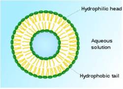

Figure 1. Liposome structure with possible modifications and applications.
Reference: http://en.wikipedia.org/wiki/Liposome
Iodixanol which is used as a contrast agent for CT imaging has 2 disadvantages when given intravenously to a living body first is that its in vivo half life is too short and the other is its toxicity to the body. This problem provides a way to use liposomes, the amphiphilic phospholipids self assembled into vesicular structures to be used as the delivery systems for the contrast agents. Liposomes have also been explored as the vehicles for the encapsulation of imaging contrast agent, including the iodinated agent iohexol, which is used in CT for the imaging of atherosclerotic plaques. The liposomal encapsulation provides a promising alternative to the traditional bolus injections of contrast media that are currently used in CT angiography by increasing the contrast agent’s in vivo half life against immune system while reducing its toxicity as compared to free, intravenously injected agent.
Experiment
In this work, the liposomal bilayer made of phospholipids DPPC, DPPE and cholesterol, covered by polyethylene glycol for increasing its stability in vivo, have been used as carrier for the contrast agent iodixanol. The liposome film formed using above encapsulates iodixanol as contrast agent and is then extruded to form 100 nm liposomes. Following this the liposomes are checked for their efficiency as a contrast agents through CT imaging. After determining the efficiency of liposomes, they are tested for in vivo imaging. The liposome sample is injected in a mouse in its eye vein to determine whether the vascular system is visible or not through the contrast agent in the liposomes.
Results
The results of in vitro and in vivo imaging of iodinated liposomes shows up considerable increase in the amount of iodixanol, encapsulated within the liposomes, per weight of the lipids in the sample and the liposome sample makes the vascular system visible.
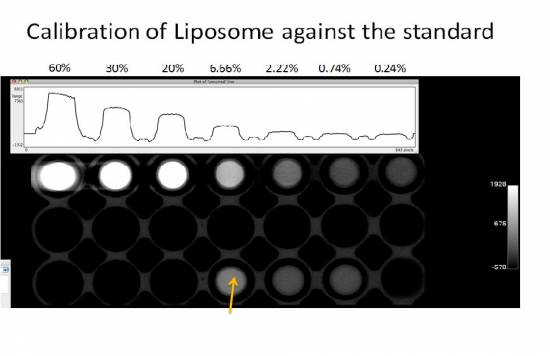 Figure 2. Liposome(marked by arrow)calibrated against standard of Iodixanol.

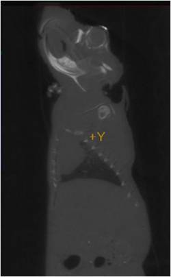
Figure 3. Left and right sagittal CT images of mouse with iodinated liposome injected in left eye vein
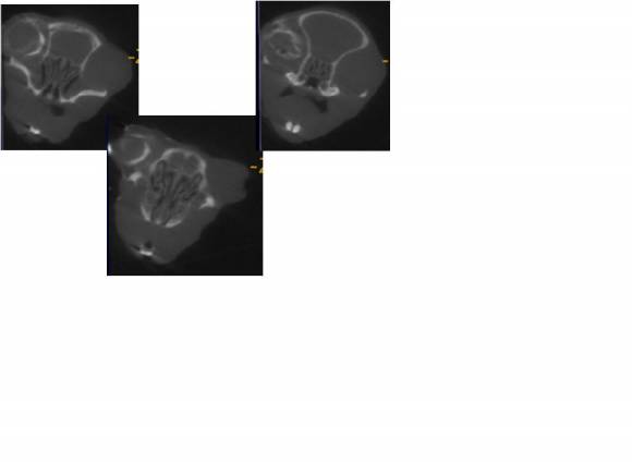 Figure 4. Transverse CT image of mouse
Latest results of in vitro and in vivo imaging of liposomes sample.

Figure 5. Comparison of CT image of liposome sample(green arrow) to a bone as a standard(blue arrow)

Figure 6. Plot of calibration of liposome contrast(green dot) against a standard of bone(blue dot).

Figure 7. CT image of liposomes injected intravenously in a 4 days old chick that makes the it visible.

Figure 8. CT image of liposomes injected intravenously in a 4 days old chick that makes the embryo and vitelline veins around visible.
Future Work
This includes determination of size and structure of liposomes, further increase in the encapsulation efficiency of liposomes and targeting cancer cells with the contrast agent.
Tanwi Kaushik
tk443@cornell.edu